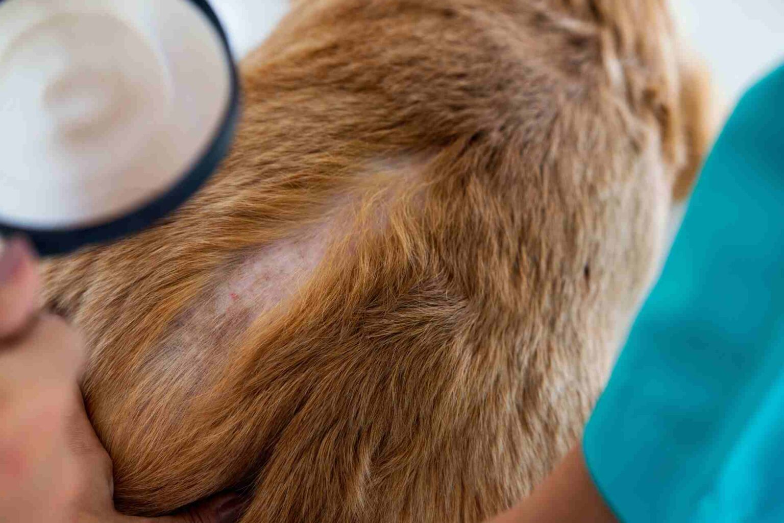
Dogs with atopic dermatitis show, similarly to humans, an altered lipid profile in the areas affected by the condition. For example, the expression of free fatty acids and certain subclasses of ceramides was found to be reduced. It would thus seem to confirm the role of lipids in maintaining the integrity of the skin barrier and, consequently, in the development of atopic dermatitis.
This is shown in a study by Suttiwee Chermprapai and colleagues from Utrecht University (Netherlands), recently published in BBA- Biomembranes.
Atopic dermatitis (AD) is a condition characterized by a genetic predisposition to chronic inflammation and severe itching. However, the pathogenesis in dogs is not yet fully known. However, dysfunction of the skin barrier, particularly the stratum corneum, could facilitate penetration of allergens resulting in immune activation and inflammation. Further alteration of the local microbiota could then aggravate the clinical picture.
Considering the composition of the epidermis, the lipid component plays a key role in its structure and function. Indeed, clinical studies of subjects with AD have shown profound qualitative alteration in the lipidome of affected areas compared with healthy controls. Based on these results, the researchers then wanted to examine the lipid component in dogs on dogs. To do so, axillary and groin skin tissue samples were taken from five dogs with AD and three healthy controls. Below are the results obtained after lipid extraction and analysis by X-ray and mass spectrometry:
- the lamellar organization of the stratum corneum in the AD group showed high variation compared with controls
- no difference was found in the relative abundance of total cholesterol and ceramides between the two groups. In contrast, significantly lower expression of free fatty acids (FFA) was recorded in dogs with AD (9.6 ± 1.4% vs. 13.9 ± 1.0%)
- subclasses of ceramides CER[EOS], CER[NS/NdS] and CER[AS/NH] were found to be the predominant ones overall. Among them, however, the ratio of CER lipids[NS] C44/C34 (i.e., with 44 or 34 carbons atoms) was found to be decreased in the AD group. No difference, on the other hand, in the CER ratio[NdS] C44/C34 between the groups
- the CER report[NS] C44/C34 showed a decreasing (nonlinear) relationship with disease severity indicated by the CADESI score. In fact, a lower C44/C34 ratio (5-25) was found in dogs with moderate-severe AD than in controls. This ratio was even lower in the most severe specimens (0-12).
Dogs with atopic dermatitis would then have an altered skin lipid profile in structure and expression. Disease severity would then appear to correlate with levels of ceramides with 34 carbons atoms. Given the small sample size, however, further investigation into the lipid role of this disease in dogs is needed.


